Craniopharyngiomas
Craniopharyngiomas are slow-growing and benign tumors that develop along the pituitary stalk, which connects the pituitary gland to the brain. While benign, these localized tumors may reach a large size before they are diagnosed and can involve the surrounding pituitary gland, third ventricle, hypothalamus and optic nerves.

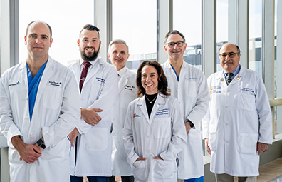
Meet Our Brain Tumor Care Team
Meet the specialists at the Gerald J. Glasser Brain Tumor Center who are experts in treating craniopharyngiomas.
Meet the TeamSymptoms
As the tumor grows, it puts pressure on the surrounding parts of the brain. This causes symptoms, which vary depending on the area it is compressing.
- Pituitary gland – causes hormone deficiency and related developmental delay, short stature, obesity and a range of other hormonal problems, including the inability to regulate water balance.
- Optic nerves – may lead to visual deficits, usually partial blindness in the outer half of the right and left peripheral vision (bitemporal hemianopsia).
Larger tumors can also cause headaches and nausea due to increased pressure within the skull or a backup of the cerebrospinal fluid that surrounds the brain and spinal cord (hydrocephalus).
Diagnosis
Craniopharyngiomas are diagnosed through a physical exam and specialized testing, including:
- Endocrine assessment – Blood tests are used to evaluate the patient’s hormone levels and determine if the pituitary gland is functioning normally. An endocrinologist may be consulted at this stage.
- Ophthalmic assessment – An ophthalmologist may examine the patient’s eyes and perform a visual field test to determine if the tumor is affecting clarity of vision or impairing peripheral vision.
- Imaging studies – Magnetic resonance imaging (MRI) and computed tomography (CT) scans with contrast dye determine the size, location and borders of the tumor.
An exact diagnosis can only be confirmed once the tumor has been removed. Tissue analysis by a neuropathologist defines the diagnosis and genetic profile of the tumor.
Treatment
Surgery is the initial treatment for a craniopharyngioma. Our team will develop a personalized plan to remove as much of the tumor as safely possible. However, craniopharyngiomas can adhere to structures within the skull base, so complementary follow-up treatments are sometimes used instead of complete removal.
We use the latest technology for precision during surgery, like high-powered microscopes, ultrasonic aspirators and stereotactic image-guided techniques.
Surgical procedures include:
- Minimally invasive endoscopic surgery – usually performed through the nasal sinuses by a neurosurgeon and otolaryngologist (ENT) who are experts in skull base surgery.
- Orbitozygomatic craniotomy – an advanced technique for skull base surgery where the bone near the cheek and eye is temporarily removed. This allows the surgeon to perform microsurgery in hard-to-reach areas of the brain and avoid unnecessary brain manipulation and risk of harm.
The diagnosis and molecular profile of each tumor is individually reviewed by our multi-disciplinary tumor board. Together, our experts recommend the best personalized and targeted treatment options incorporating the latest molecular diagnostics, treatment protocols and participation in national clinical trials. Follow-up treatments can include:
- Observation – Even when the tumor has been totally removed, surveillance MRI or CT scans are used to watch for recurrence.
- Stereotactic radiosurgery (CyberKnife®) – This non-invasive treatment option is used after surgery for small or residual tumors in hard-to-reach locations or if a tumor returns after surgery.
Our team will closely monitor you and personalize your follow-up care. Our patient navigator will also connect you with our support group and other resources.